Treatment:
The treatment recommendation is based on patient’s health and severity of achalasia. The following treatments are recommended:
Endoscopic Treatment:
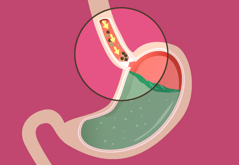
Endoscopic balloon dilatation therapy:
Once the diagnosis of achalasia is confirmed, the doctor will put down an endoscope through the mouth to the site of LES. A balloon is then placed across the sphincter, and gradually dilated with saline to a certain pressure enough to disrupt the muscle fibres of the LES. It offers temporary relief, and may need repeated attempts to give symptomatic relief. It is not a permanent solution, and the patient will end up needing a POEM or laparoscopic surgery to address it.
Per Oral Endoscopic Myomectomy (POEM):
In POEM, the surgeon carries out the procedure via the endoscope introduced through the mouth. Using specialised instruments, the surgeon makes a small cut on the inner lining of the esophagus and creates a tunnel under the mucosal layer all the way up to the tight lower esophageal sphincter. The LES is then divided using a small knife via the endoscope, and the mucosal incision in the esophagus higher above is closed with clips. The patient has a check endoscopy and dye swallowing test the following day to confirm no side effects from the procedure. The patient is discharged after 1 day and can resume normal diet. Currently, POEM has equivalent results to laparoscopic surgery, and remains scarless. There is a slightly higher risk of reflux, and the advice would be appropriately given.
Surgery: Laparoscopic Heller’s Cardiomyotomy:
This has been the ‘ gold standard of treatment’ for many years with good outcomes and resolution of symptoms. This procedure is done laparoscopically/ key hole method which gives quicker recovery. Small cuts are made on the abdomen, and surgeon uses laparoscopic instruments to perform the surgery. The surgeon makes a cut over the LES sphincter area to divide the muscle fibres only (protecting the inner lining mucosa) and follows the cut with a fundoplication procedure ( stomach wrap) to prevent acid reflux from stomach into oesophagus. The surgery usually lasts 2-3 hours and most patients are discharged within 1-2 days.
Follow up:
Post achalasia surgery ( endoscopy or surgery), the patient will require dedicated endoscopic screening every 2-3 years depending on the severity pre-procedure. Due to increased acid reflux post treatment, the patient’s are prone to develop changes in the lining in the lower esophagus leading to esophagitis, BARRETT’S esophagus and in small proportion of patients, development of cancer. Hence, endoscopic screening is the only method to keep an eye out for these developments.